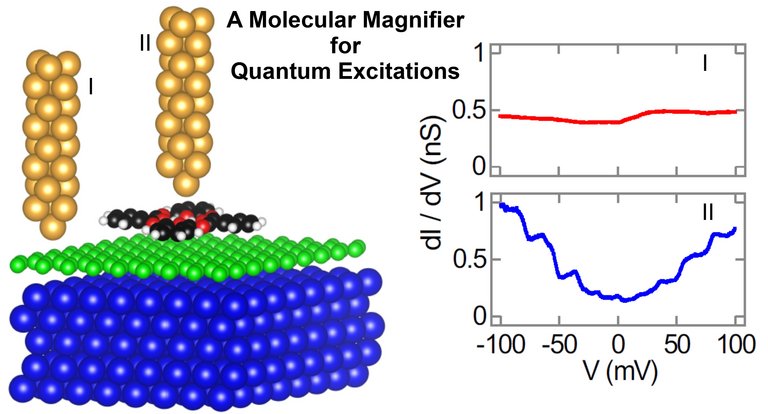
In everyday life, we use a magnifying glass to visualize objects that are difficult or even impossible to see with the naked eye. A pertinent question arises as to the magnified visualization of quantum excitations, i.e., of the transfer of smallest energy portions. Physicists from Ilmenau and Lyngby have found the answer and showed how a single molecule can magnify the spectroscopic signature of graphene vibrations, that is, the oscillatory motion of carbon atoms in the honeycomb lattice of the two-dimensional material, and how the signal of the vibrational excitation can be detected by means of a scanning tunneling microscope (STM).
In contrast to optical microscopes, which deploy lenses for imaging, an STM uses the quantum mechanical tunneling current, which is injected from an atomically sharp tip into the sample and, thereby, passes through the vacuum barrier between tip and sample, which for a classical current would be impossible. Owing to the local injection of the current and its exponential dependence on the tip-surface distance the STM is capable of atomic resolution, i.e., atoms of surfaces or adsorbed molecules can be imaged. Moreover, the STM represents a tool that enables the manipulation of matter atom by atom, which provides a direct connection between the macroscopic world and the nanocosmos.
The information on quantum excitations, which play the central role in the present studies, can be extracted from current-voltage characteristics acquired atop deliberate surface sites: the applied bias voltage provides energy for the tunneling electrons that can be transferred to, e.g., atom vibrations of the graphene lattice – a process that is referred to as inelastic electron tunneling. Upon energy transfer the current changes at a specific bias voltage. By means of phase-sensitive detectors the minute energy transfer can be measured.


